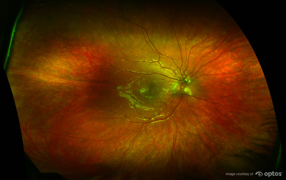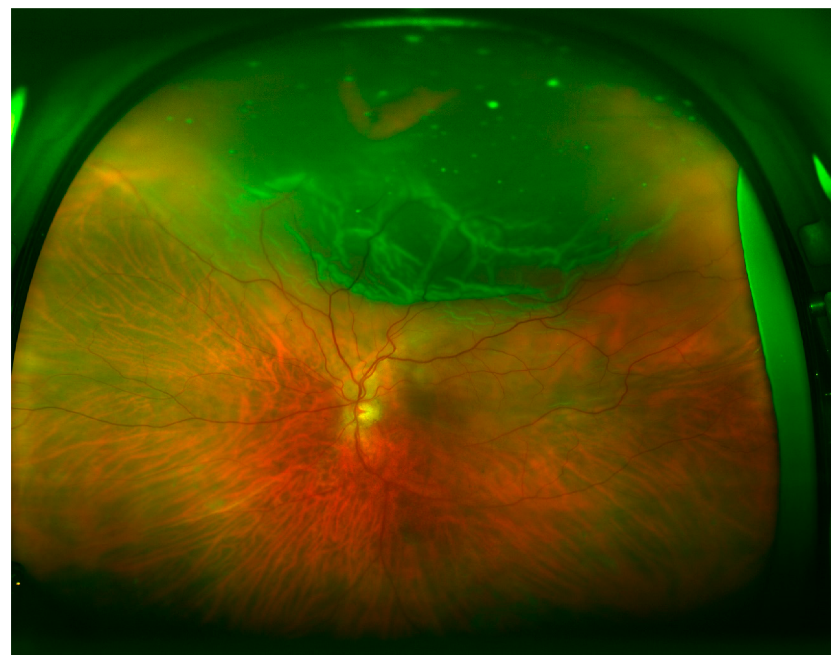
Simultaneous, non-contact, central pole-to-periphery views of up to 82% or 200 degrees of the retina (back of eye) are displayed in one single capture, compared to 45 degrees achieved with conventional methods. With the optomap technology, the Optos retinal scanner can take a 200° panoramic digital image that’s about 80 of your eye’s fundus It equates to a 50 increase over the next closest imaging device. These conditions may otherwise go undetected using traditional examination techniques and equipment. In fact, they only capture between 10° to 100° of the fundus or the interior part of the eye. Optos’ patented ultra-widefield digital laser scanning technology acquires images that support the detection, diagnosis, analysis, documentation, and management of ocular pathology and systemic disease that may first present in the periphery of the retina (back of eye). Distressed by the diagnostic methods available, Leif’s father, Douglas, designed the Optomap retinal exam.

He was getting regular eye exams, but conventional tests available at the time made a thorough examination difficult. The Optomap story: In 1990, 5 year old Leif Anderson went blind in one eye due to an undiagnosed retinal detachment. The exam is quick, painless, and may not require dilation drops. This can lead to early detection of common diseases, such as glaucoma, diabetes, macular degeneration, and even cancer. Your doctor offers optomap to patients of all ages. The ultra-widefield AF images were obtained using an Optomap P200Tx fundus camera (Optos, Dunfermline, UK) from both eyes during follow-up visits. Many conditions, such as retinal detachments and retinal holes can be treated successfully if caught early. While eye exams generally include a look at the front of the eye to evaluate health and prescription changes, a thorough screening of the retina is critical to verify that your eye is healthy. Autofluorescence Imaging in the Long-Term Follow-Up of Scleral Buckling Surgery for Retinal Detachment. In addition, many vision problems begin at an early age, so it’s important for children to receive proper eye care from the time they are infants. Today, a whole range of eye problems can be treated successfully without total vision loss. Regular eye care and exams can protect and prevent many eye diseases, if detected early. We all want to protect our eyesight and overall health for ourselves and our family – that is why annual eye exams are important.
#Retinal detachment optomap archive#
No adverse health effects have been reported in over 150 million sessions.Optomap Retinal Imaging | Miss-Lou Eye Care alarm-ringing ambulance angle2 archive arrow-down arrow-left arrow-right arrow-up at-sign baby baby2 bag binoculars book-open book2 bookmark2 bubble calendar-check calendar-empty camera2 cart chart-growth check chevron-down chevron-left chevron-right chevron-up circle-minus circle city clapboard-play clipboard-empty clipboard-text clock clock2 cloud-download cloud-windy cloud clubs cog cross crown cube youtube diamond4 diamonds drop-crossed drop2 earth ellipsis envelope-open envelope exclamation eye-dropper eye facebook file-empty fire flag2 flare foursquare gift glasses google graph hammer-wrench heart-pulse heart home instagram joystick lamp layers lifebuoy link linkedin list lock magic-wand map-marker map medal-empty menu microscope minus moon mustache-glasses paper-plane paperclip papers pen pencil pie-chart pinterest plus-circle plus power printer pushpin question rain reading receipt recycle reminder sad shield-check smartphone smile soccer spades speed-medium spotlights star-empty star-half star store sun-glasses sun tag telephone thumbs-down thumbs-up tree tumblr twitter user users wheelchair write yelp youtubeĭownload a brochure for more information. Optomap images are created by non-invasive, low-intensity scanning lasers. However, it is generally recommended that you have an optomap each time you have an eye exam.

This is a decision that should be made by your doctor.
:max_bytes(150000):strip_icc()/GettyImages-308783-003-56acdcd85f9b58b7d00ac8e8.jpg)

Unlike traditional retinal exams, the optomap image can be saved for future comparisons. The ultra-widefield optomap may help your eye doctor detect problems more quickly and easily. It is completely comfortable and the scan takes less than a second. It is the only technology that can capture 82% view of your retina at one time. The optomap is a digital image of the retina produced by Optos scanning laser technology. retinal easily twenty years ago when his then 5 year old son experienced permanent vision loss from an undetected retinal detachment. In fact, many vision problems begin in early childhood, so it's important for children to receive quality routine eye care. Some of the first signs of diseases such as stroke, diabetes and even some cancers can be seen in your retina, often before you have other symptoms. Frequently Asked Questions about an optomap


 0 kommentar(er)
